A vestigial organ, by evolutionary definition, is an
organ that was once useful during a previous stage of your evolution
for a supposed ancestor, but in the course of time, that organ was no
longer needed, but continued to remain in the body.
Analysis:
If vestgial organs would prove anything, they prove devolution, NOT evolution !
Darwinists claim that some of our organs are falling into disuse. Yet, in contrast, they provide us with no one NEW, developing organ. The "vestigial organs" idea, if it could be true, would only prove the opposite: devolution!
2- Not for survival !
Not all organs are necessary for survival,this doesn't mean they are useless!
You have 2 lungs, you need one to survive. You have 2 kidneys, you need 1/10 for survival. No one claimed the other lung may be 'just for fun'.
you will survive if your eyes and arms are cut out, and they are not "vestigial," or useless organs.
3- Causing problems ?!
people have far more problems with their lungs, hearts and stomachs, than they have with 'vestigial' organs. Almost any organ in your body can kill you if is it sufficiently diseased. How many people die of heart attacks vs. appendicitis? The heart, the physical or the spiritual one, is far more troublesome. If your lungs become infected, you can die but no one suggests removing the lungs as a preventive measure during surgery for another reason.
4- Affirming the consequent !
[If we ignored the fact that they actually prove devolution] For this type of argument to be used, one has to assume evolution to be true in the first place! So, if vestigial organs are obviously left over from our evolutionary heritage, then evolution must be true. Here evolution is assumed to be true in order to make the argument. This is a logical fallacy.
5 - FOUNDED ON IGNORANCE:
How did such an idea become accepted in the first place? It happened in a time of great ignorance. The whole idea of "vestigial organs" was originally conceived back in the early 1800s, at a time when physicians were still blood-letting in order to cure people of infection. But since that time there has been an immense quantity of research in every imaginable field. There is now no doubt by competent biologists that every large and small part of the human body has a special function during the life of the individual.
It strongly appears that the true "vestigial organ" in earlier times, was an ignorant mind; a mind that did not know why organs were in the body, and was too impatient and lazy to do the laborious work needed to identify functions. But we should not want to call ignorance a proof of evolution.
Just because someone doesn’t know the function of something, that does not mean there is none, which means:If none of our organs nor genes were "known" to have function, this wouldn't prove it's vestigial, It's more appropriate to say: Human don't know Yet!
http://en.wikipedia....ert_Wiedersheim
Blechschmidt notes that
Reputable scientists now recognize that the evolutionary teaching of "vestigial organs" actually retarded scientific knowledge for decades. Instead of finding out what the appendix was for, it was called "vestigial" and was cut out. Researchers were told it was a waste of time to study any possible use for it. For the same reason, lots of children have had their tonsils removed, when they really needed them!
"The existence of functionless 'vestigial organs' was presented by Darwin, and is often aced by current biology textbooks, as part of the evidence for evolution . . An analysis of the difficulties in unambiguously identifying functionless structures . . leads to the conclusion that 'vestigial organs' pride no evidence for evolutionary theory." *S.R. Scudding, "Do 'Vestigial Organs' Provide Evidence for Evolution?" Evolutionary Theory, Vol. (May 1961), p. 394.
7- Lost primary functions !
Confronted with the fact that many previously thought to be "vestigial" organs, are now known to have functions , Darwinists made a funny conjecture that these organs "developed secondary functions", however, they didn't provide the scientific criteria to determine if a function is primary or secondary !!
8- Some known functions of some "alleged vestigial organs":
Appendix:
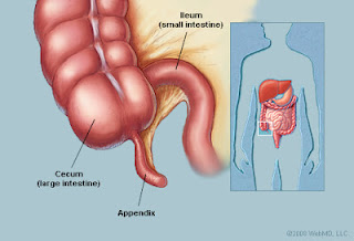
- The appendix is now known to be an important part of what is called the reticulo-enadothelial system of the body. Like the tonsils, the appendix fights infection.
There are collections of lymphocytes (body defence cells) in its lining.
The appendix is part of a system that determines which microbes are allowed to live in the intestines and which ones are not. The large intestine needs to have a healthy population of harmless bacteria living on its inner surface.
Babies are not born with these microbes. Babies develop in a germ-free environment in their mother’s womb so during infancy and childhood the immune system has to learn which microbes can live on the body surfaces and which cannot. Even the good microbes need to be kept in their place and your immune system helps keep them there throughout your life.
- When the large bowel becomes inflamed and its population of good bacteria is lost due to outpouring fluid that is part of the inflammatory response to injury and infection, the appendix acts as a “safe house” for good bacteria, which can then repopulate the large bowel when the inflammation is over. For more information on this function see article in Science Daily, 8 Oct 2007.
- One study done by Dr. Howard R. Bierman on hundreds of patients with leukemia, Hodgkin’s disease, cancer of the colon, and cancer of the ovaries showed that 84% of these patients had their appendix removed, while in a healthy control group only 25% had it removed. [Bergman and Howe, p. 45] This is a positive correlation, indicating a possible role of the appendix in preventing these diseases.
“Maybe it's time to correct the textbooks,” said researcher William Parker, an immunologist at Duke University Medical Center in Durham, NC “Many biology texts today still refer to the appendix as a 'vestigial organ.”
The Coccyx:
Another organ declared useless by evolutionists is the coccygeal vertebrae (the coccyx). This is the bottom of your spine.The fact that the coccyx is made of several segments fused together is no indication that it used to be a mobile tail.
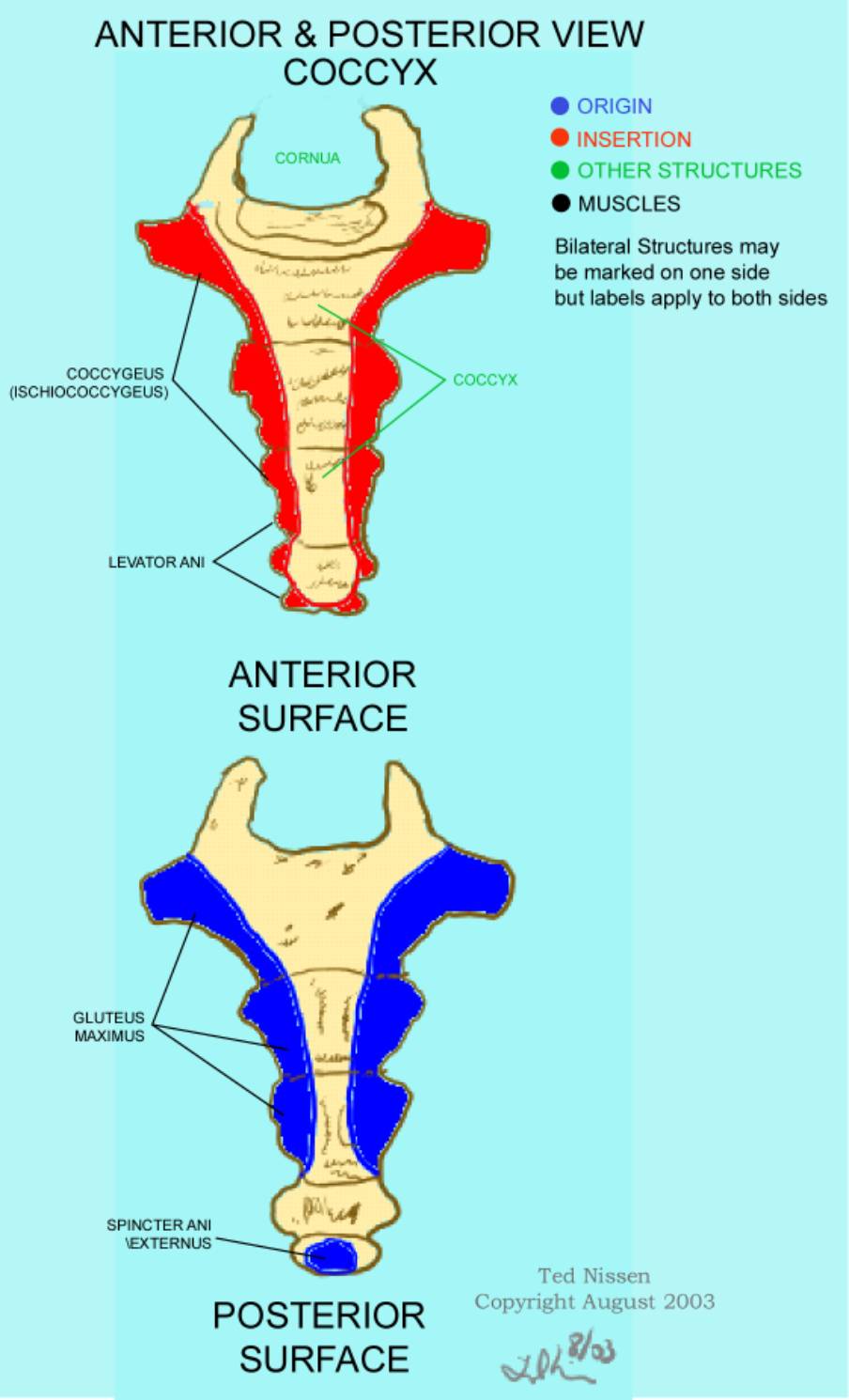 The segmented structure allows it to grow
during foetal development and childhood. Scientists have found that
important muscles attach to those bones. The pelvic floor muscles and ligaments help with maintaining upright stance and walking, and support the internal organs of pelvic cavity.
The segmented structure allows it to grow
during foetal development and childhood. Scientists have found that
important muscles attach to those bones. The pelvic floor muscles and ligaments help with maintaining upright stance and walking, and support the internal organs of pelvic cavity.
http://www.laserspin...natomy/tailbone
http://education.yah...ects/subject/24
http://education.yah...cts/subject/213
http://www.angelfire...s/tailbone.html
Plica semilunaris of conjunctiva
Wisdom teeth:
First of all, a tooth, just like any other molar tooths has an obvious function, namely to grind food. We have 32 teeth, including these wisodm teeth. Of course we could still grind food without our 4 wisdom teeth. We could probably also be able to grind food with 26, 24 or with 20 teeth. Be that as it may, the wisdom teeth are obviously not vestigal. It has also been suggested, that earlier generations of humans which ate food that was allot rougher, and didn't have the same protection and care wore down their teeth a lot faster, therefor, a new set of teeth, at the age of 20 to 25 was very practical for them.
Opposition to Prophylactic Removal of Third Molars (Wisdom Teeth)
http://www.apha.org/...ult.htm?id=1371
The Vomeronasal (Jacobson’s) Organ

Erector pili:
Related: Why Mammal Body Hair Is an Evolutionary Enigma
http://faculty.washi...r/receptor.html
http://www.keratin.com/aa/aa031.shtml
http://jap.physiolog...6/2/256.extract
http://www.scientifi...ile-sl-11-12-13
Plantaris
In human beings it is tiny and may be absent (7–20% of population)(Simpson SL, Hertzog MS, Barja RH. The plantaris tendon graft: an ultrasound study. J Hand Surg [Am] 1991;16:708–711)

In humans, Plantaris aids to:
Analysis:
1- Devolution Not evolution !
If vestgial organs would prove anything, they prove devolution, NOT evolution !
Darwinists claim that some of our organs are falling into disuse. Yet, in contrast, they provide us with no one NEW, developing organ. The "vestigial organs" idea, if it could be true, would only prove the opposite: devolution!
2- Not for survival !
Not all organs are necessary for survival,this doesn't mean they are useless!
You have 2 lungs, you need one to survive. You have 2 kidneys, you need 1/10 for survival. No one claimed the other lung may be 'just for fun'.
you will survive if your eyes and arms are cut out, and they are not "vestigial," or useless organs.
3- Causing problems ?!
people have far more problems with their lungs, hearts and stomachs, than they have with 'vestigial' organs. Almost any organ in your body can kill you if is it sufficiently diseased. How many people die of heart attacks vs. appendicitis? The heart, the physical or the spiritual one, is far more troublesome. If your lungs become infected, you can die but no one suggests removing the lungs as a preventive measure during surgery for another reason.
4- Affirming the consequent !
[If we ignored the fact that they actually prove devolution] For this type of argument to be used, one has to assume evolution to be true in the first place! So, if vestigial organs are obviously left over from our evolutionary heritage, then evolution must be true. Here evolution is assumed to be true in order to make the argument. This is a logical fallacy.
5 - FOUNDED ON IGNORANCE:
How did such an idea become accepted in the first place? It happened in a time of great ignorance. The whole idea of "vestigial organs" was originally conceived back in the early 1800s, at a time when physicians were still blood-letting in order to cure people of infection. But since that time there has been an immense quantity of research in every imaginable field. There is now no doubt by competent biologists that every large and small part of the human body has a special function during the life of the individual.
It strongly appears that the true "vestigial organ" in earlier times, was an ignorant mind; a mind that did not know why organs were in the body, and was too impatient and lazy to do the laborious work needed to identify functions. But we should not want to call ignorance a proof of evolution.
Just because someone doesn’t know the function of something, that does not mean there is none, which means:If none of our organs nor genes were "known" to have function, this wouldn't prove it's vestigial, It's more appropriate to say: Human don't know Yet!
http://en.wikipedia....ert_Wiedersheim
Blechschmidt notes that
"no organ could exist that is functionless during its development," an axiom that also applies to the nervous system.(Blechschmidt, The Ontogenetic Basis of Human Anatomy, 91. )
6- HINDERS SCIENCEReputable scientists now recognize that the evolutionary teaching of "vestigial organs" actually retarded scientific knowledge for decades. Instead of finding out what the appendix was for, it was called "vestigial" and was cut out. Researchers were told it was a waste of time to study any possible use for it. For the same reason, lots of children have had their tonsils removed, when they really needed them!
"The existence of functionless 'vestigial organs' was presented by Darwin, and is often aced by current biology textbooks, as part of the evidence for evolution . . An analysis of the difficulties in unambiguously identifying functionless structures . . leads to the conclusion that 'vestigial organs' pride no evidence for evolutionary theory." *S.R. Scudding, "Do 'Vestigial Organs' Provide Evidence for Evolution?" Evolutionary Theory, Vol. (May 1961), p. 394.
7- Lost primary functions !
Confronted with the fact that many previously thought to be "vestigial" organs, are now known to have functions , Darwinists made a funny conjecture that these organs "developed secondary functions", however, they didn't provide the scientific criteria to determine if a function is primary or secondary !!
8- Some known functions of some "alleged vestigial organs":
Appendix:
The
appendix is a small blind-ended tubular structure attached to the large
intestine close to where it joins the small intestine. It has no
digestive function and is commonly assumed to a vestigial organ left
behind from a plant-eating ancestor.
Evolutionists have postulated that in the past, man had a larger cecum, but as man progressed from a higher-fiber diet to a lower-fiber diet, the larger cecum became less necessary. Thus the appendix is said to have resulted from a loss of cecal size.
Evolutionists have postulated that in the past, man had a larger cecum, but as man progressed from a higher-fiber diet to a lower-fiber diet, the larger cecum became less necessary. Thus the appendix is said to have resulted from a loss of cecal size.
- The appendix is now known to be an important part of what is called the reticulo-enadothelial system of the body. Like the tonsils, the appendix fights infection.
There are collections of lymphocytes (body defence cells) in its lining.
The appendix is part of a system that determines which microbes are allowed to live in the intestines and which ones are not. The large intestine needs to have a healthy population of harmless bacteria living on its inner surface.
Babies are not born with these microbes. Babies develop in a germ-free environment in their mother’s womb so during infancy and childhood the immune system has to learn which microbes can live on the body surfaces and which cannot. Even the good microbes need to be kept in their place and your immune system helps keep them there throughout your life.
- When the large bowel becomes inflamed and its population of good bacteria is lost due to outpouring fluid that is part of the inflammatory response to injury and infection, the appendix acts as a “safe house” for good bacteria, which can then repopulate the large bowel when the inflammation is over. For more information on this function see article in Science Daily, 8 Oct 2007.
- One study done by Dr. Howard R. Bierman on hundreds of patients with leukemia, Hodgkin’s disease, cancer of the colon, and cancer of the ovaries showed that 84% of these patients had their appendix removed, while in a healthy control group only 25% had it removed. [Bergman and Howe, p. 45] This is a positive correlation, indicating a possible role of the appendix in preventing these diseases.
- Your Appendix Could Save Your Life
http://blogs.scienti...save-your-life/
http://www.scientifi...e-function-of-t
http://www.ncbi.nlm....pubmed/15228837
http://www.ncbi.nlm....pubmed/17936308
http://www.ncbi.nlm....pubmed/21370495
http://www.sciencedi...02251930700416X
http://www.ncbi.nlm....les/PMC3279496/
http://www.livescien...-promising.html
Scientists Finally Discover The Function of the Human Appendix
“Maybe it's time to correct the textbooks,” said researcher William Parker, an immunologist at Duke University Medical Center in Durham, NC “Many biology texts today still refer to the appendix as a 'vestigial organ.”
The Coccyx:
Another organ declared useless by evolutionists is the coccygeal vertebrae (the coccyx). This is the bottom of your spine.The fact that the coccyx is made of several segments fused together is no indication that it used to be a mobile tail.
http://www.laserspin...natomy/tailbone
http://education.yah...ects/subject/24
http://education.yah...cts/subject/213
http://www.angelfire...s/tailbone.html
Plica semilunaris of conjunctiva
The plica semilunaris is a small fold of bulbar conjunctiva on the medial canthus of the eye.

It functions during movement of the eye, to help maintain tear drainage via the lacrimal lake, and to permit greater rotation of the globe, for without the plica the conjunctiva would attach directly to the eyeball, restricting movement.
It functions during movement of the eye, to help maintain tear drainage via the lacrimal lake, and to permit greater rotation of the globe, for without the plica the conjunctiva would attach directly to the eyeball, restricting movement.
"Four
fornices are formed by the conjunctiva where it is reflected from the
eyelids to the globe. The superior fornix is the largest in both size
and volume. It is formed and maintained by fine smooth muscle slips that
pass from the levator palpebral superioris muscle and insert into the
conjunctiva. This attachment prevents the conjunctiva from folding
downward over the cornea. It also prevents the fornix from developing
sags as the globe moves upward, which might obstruct vision. The
temporal fornix is attached by fine fibrous slips to the tendon of the
lateral rectus muscle, again maintaining the relative position of the
fornix during horizontal movements of the globe. The inferior fornix is
attached to the tendon of the inferior rectus, which prevents its
movement. The plica semilunaris performs the same function as a fornix
medially, and is a reversed fornix with the fold of conjunctiva lying
externally (Fig. 3). During a medial gaze, the fornix has a variable
depth. This occurs because the fibrous slips that link the conjunctiva
to the tendon of the medial rectus muscle insert onto the deep surface
of the plica and caruncle. Contraction of the medial rectus tightens
these slips, forming a cul-de-sac medially as the globe adducts. On
maximal medial rotation, the plica partially unfolds to form a true
fornix similar to that present in other areas. (Fig. 3). During a medial
gaze, the fornix has a variable depth. This occurs because the fibrous
slips that link the conjunctiva to the tendon of the medial rectus
muscle insert onto the deep
surface of the plica and caruncle. Contraction of the medial rectus tightens these slips, forming a cul-de-sac medially as the globe adducts. On maximal medial rotation, the plica partially unfolds to form a true fornix similar to that present in other areas."(Dartt, Darlene A. (2006). "The Conjunctiva—Structure and Function". Duane's Foundations of Clinical Ophthalmology 2. Philadelphia: Lippincott Williams & Wilkins. Chapter 2)
surface of the plica and caruncle. Contraction of the medial rectus tightens these slips, forming a cul-de-sac medially as the globe adducts. On maximal medial rotation, the plica partially unfolds to form a true fornix similar to that present in other areas."(Dartt, Darlene A. (2006). "The Conjunctiva—Structure and Function". Duane's Foundations of Clinical Ophthalmology 2. Philadelphia: Lippincott Williams & Wilkins. Chapter 2)
http://www.oculist.n.../v8/v8c002.html
The relationship between the plica semilunaris and caruncle and the bulbar conjunctiva, eyelids, and lacrimal puncta is important in several ways. Any change in these structures due to scarring or other fibrous changes could mechanically limit rotation of the globe. In addition, keratinization, hypertrophy, or retraction of the caruncle may interfere with mucus and foreign body excretion, resulting in dysfunction of the lacrimal drainage system.
N.B:
Darwinists say it's is the vestigial non-functional remnant of the the "third eyelid"(nictitating membrane) which is drawn across the eye for protection in some animals e.g:camels, sharks & birds.
Wisdom teeth:
First of all, a tooth, just like any other molar tooths has an obvious function, namely to grind food. We have 32 teeth, including these wisodm teeth. Of course we could still grind food without our 4 wisdom teeth. We could probably also be able to grind food with 26, 24 or with 20 teeth. Be that as it may, the wisdom teeth are obviously not vestigal. It has also been suggested, that earlier generations of humans which ate food that was allot rougher, and didn't have the same protection and care wore down their teeth a lot faster, therefor, a new set of teeth, at the age of 20 to 25 was very practical for them.
Opposition to Prophylactic Removal of Third Molars (Wisdom Teeth)
http://www.apha.org/...ult.htm?id=1371
The Vomeronasal (Jacobson’s) Organ
The adult human VNO displays
species-specific, gender-dimorphic and highly stereospecific responses
to ligands. The organ's local response, or electrovomerogram, is
followed by gender-specific behavioral changes, modulation of autonomic
nervous system function, or the release of gonadotropins from the
pituitary gland. Functional brain imaging studies revealed consistent
activation of the hypothalamus, amygdala and cingulate gyrus-related
structures during adult human VNO stimulation. These findings present
new information supportive of a functional vomeronasal system in adult
humans.
http://www.ncbi.nlm..../pubmed/9929629
These data demonstrate the existence of a functional vomeronasal-pituitary pathway in adult humans. In addition to the effect on gonadotropin pulsatility, the vomeropherin also produces concurrent reflex autonomic effects after VNO stimulation. These included decreased respiratory frequency, increased cardiac frequency, and event-related changes of electrodermal activity and EEG pattern. Therefore, this investigation also provides evidence for functional connections between the VNO and a variety of hypothalamic areas in adult humans.
http://www.ncbi.nlm..../pubmed/8836161
Recent progress in the neurobiology of the vomeronasal organ.
http://www.ncbi.nlm....pubmed/12203701
The researchers believe that the cells in the VNO send electrical impulses to the hypothalamus which stimulates the pituitary gland to release or stop releasing certain hormones (Lawton 1997).
http://www.macaleste...attraction.html
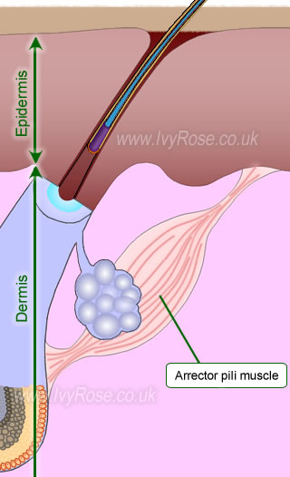
Erector pili:
Piloerection refers to a reaction of the
sympathetic nervous system that causes hair follicles to protrude
outwards from the skin. Commonly called goosebumps or gooseflesh, it is a
physiological response to cold air and intense emotions, especially
fear. It is most common to see goosebumps on the forearms, though they
can also appear on the legs, buttocks, chest, and neck on some people.
Another medical term for piloerection is cutis anserina. Goosebumps appear when the sympathetic nervous system causes the arrectores pilorum muscles under the skin to contract.
Goosebumps may help to conserve heat when you're exposed to cold. They may do this in several ways:
Another medical term for piloerection is cutis anserina. Goosebumps appear when the sympathetic nervous system causes the arrectores pilorum muscles under the skin to contract.
Goosebumps may help to conserve heat when you're exposed to cold. They may do this in several ways:
- As with larger muscles, contraction of the muscles in the skin (called "arrectores pilorum") makes heat.
- The raised hair follicles cause skin pores to close.
- Hairs standing up trap a layer of air near the skin, holding onto body heat.
They also have an important role in keeping the skin’s oil glands unblocked.
http://books.google....ctor pili human
Quantitative pilomotor axon reflex test: a novel test:The quantitative pilomotor axon reflex test (QPART) may complement other measures of cutaneous autonomic nerve fiber function.
http://www.ncbi.nlm....pubmed/22868966
Relation to diseases:
Though rare, goosebumps can be a sign of a seizure disorder (called temporal lobe epilepsy), a disorder of the sympathetic nervous system, or a brain tumor. They are also common during heroin or other opiate withdrawal. In fact, the term "quitting cold turkey" refers to the presence of goose bumps (that mimic cold turkey flesh) during withdrawal from heroin. (The term could also have come from the expression, "talk turkey," meaning to speak bluntly or directly, without preparation.)
http://www.intelihea...d=dmtHMSContent
Goose bumps may be a kind of "skin orgasm"
http://www.healthgui...oose-Bumps.html
N.B:
Cat's body, for example, initiates piloerection to make the hairs on the back and tail stand on end. This process makes the cat appear larger than it actually is.
Piloerection is also beneficial for animals when they are exposed to extremely cold weather. Goosebumps extend the hairs on a cold animal, creating a thicker coat that offers more protection. Erected hairs provide extra insulation as they trap heat more effectively than a fur coat in its normal state.
Because piloerction in man doesn't make predators think twice before attacking & its role in heat regulation is less that animals, Darwinists declared it a useless leftover from our imaginary animal ancestors.
-----------------------http://books.google....ctor pili human
Quantitative pilomotor axon reflex test: a novel test:The quantitative pilomotor axon reflex test (QPART) may complement other measures of cutaneous autonomic nerve fiber function.
http://www.ncbi.nlm....pubmed/22868966
Relation to diseases:
Though rare, goosebumps can be a sign of a seizure disorder (called temporal lobe epilepsy), a disorder of the sympathetic nervous system, or a brain tumor. They are also common during heroin or other opiate withdrawal. In fact, the term "quitting cold turkey" refers to the presence of goose bumps (that mimic cold turkey flesh) during withdrawal from heroin. (The term could also have come from the expression, "talk turkey," meaning to speak bluntly or directly, without preparation.)
http://www.intelihea...d=dmtHMSContent
Goose bumps may be a kind of "skin orgasm"
http://www.healthgui...oose-Bumps.html
N.B:
Cat's body, for example, initiates piloerection to make the hairs on the back and tail stand on end. This process makes the cat appear larger than it actually is.
Piloerection is also beneficial for animals when they are exposed to extremely cold weather. Goosebumps extend the hairs on a cold animal, creating a thicker coat that offers more protection. Erected hairs provide extra insulation as they trap heat more effectively than a fur coat in its normal state.
Because piloerction in man doesn't make predators think twice before attacking & its role in heat regulation is less that animals, Darwinists declared it a useless leftover from our imaginary animal ancestors.
Related: Why Mammal Body Hair Is an Evolutionary Enigma
http://faculty.washi...r/receptor.html
http://www.keratin.com/aa/aa031.shtml
http://jap.physiolog...6/2/256.extract
http://www.scientifi...ile-sl-11-12-13
Plantaris
In monkeys it is connected to the toes and is useful in swinging from branches.

(Image: Plantaris in cat's leg)
(Image: Plantaris in cat's leg)
In human beings it is tiny and may be absent (7–20% of population)(Simpson SL, Hertzog MS, Barja RH. The plantaris tendon graft: an ultrasound study. J Hand Surg [Am] 1991;16:708–711)
In humans, Plantaris aids to:
- plantarflex the ankle joint
- flex the knee joint
It has been considered to be an organ of proprioceptive function for the larger, more powerful plantarflexors as it contains a high density of muscle spindles.(Moore
KL, Dalley AF, editors. Clinically Oriented Anatomy. 5. Philadelphia:
Lippincott Williams & Wilkins; 2006. pp. 648–649)
Its motor function is minimal but its long tendon can readily be harvested for plastic reconstruction elsewhere with little functional deficit.
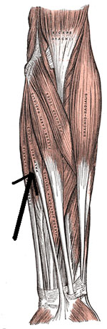
Palmaris Longus:Its motor function is minimal but its long tendon can readily be harvested for plastic reconstruction elsewhere with little functional deficit.
It's a flexor of the wrist and tenses the palmar aponeurosis & It's great candidate for plastic reconstruction.
"loss
of this tendon results in no major abnormality of hand function. But Mc
Grouther (1996) states that the main function of palmaris longus is to
anchor the skin and fascia of hand and hence to prevent degloving of
palmar skin from horizontal shearing forces. Therefore, variations in
the innervation of such a clinically important muscle should be of
interest both to the academicians and clinicians"
No comments:
Post a Comment